Womans Foot Bones Labeled On White Stock Photo Download Image Now Human Skeleton, Ankle
Anatomy The Bones Of The Foot
Because they are so complicated, human feet can be especially prone to injury. Strains, sprains, tendonitis, torn ligaments, broken bones, fallen arches, bunions, corns, and plantar warts can all occur. Here we will talk more about the anatomy of the human foot and its many moving parts.

Foot Description, Drawings, Bones, & Facts Britannica
Browse 19,300+ foot anatomy stock photos and images available, or search for foot anatomy vector to find more great stock photos and pictures. foot anatomy vector Sort by: Most popular Human foot anatomy cutaway representation, clipping path included. "Human foot anatomy cutaway representation, showing skin, veins and arterias, muscles, bones.

Foot bones anatomy Royalty Free Vector Image VectorStock
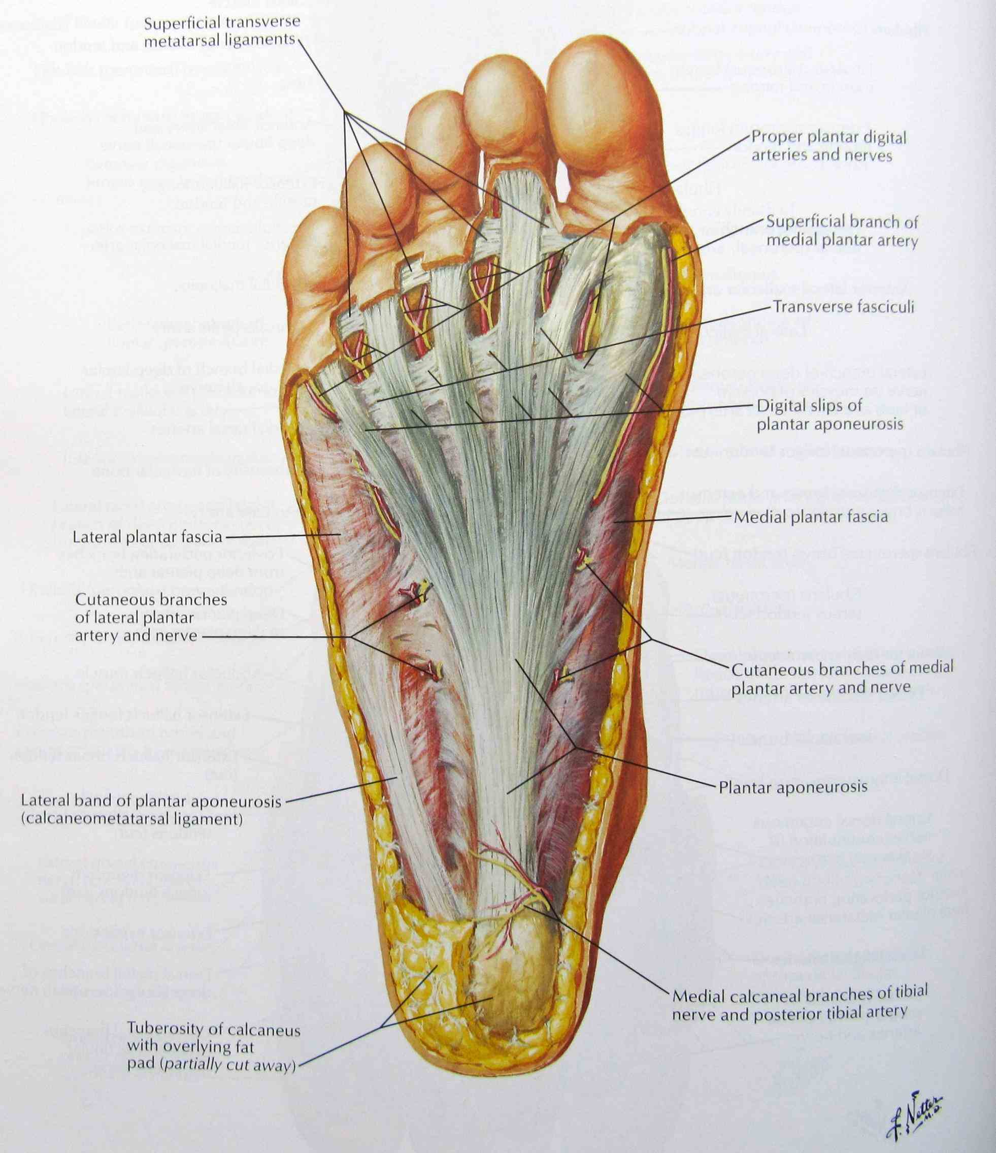
Foot: The end of the leg on which a person normally stands and walks. The foot is an extremely complex anatomic structure made up of 26 bones and 33 joints that must work together with 19 muscles and 107 ligaments to execute highly precise movements. At the same time the foot must be strong to support more than 100,000 pounds of pressure for.

3B Scientific A31L Rigid Skeletal Foot Model Portion Tibia Fibula
Skeleton Leg Icon. This 100% royalty free vector illustration features the main icon pictured in black inside a white square. The alternative color options in blue, green, yellow and red are on the right of the icon and are arranged in a vertical column. Anatomy Bones of the Feet. Orthotics for foot Superior view and.
Human Foot Bones Photograph by Pixologicstudio/science Photo Library Pixels
foot skeleton picture Like already mentioned, the hindfoot is the posterior part of the foot. Its made up of 4 bones; The calcaneus, talus, fibula, and tibia bones. 1. The tibia bone The tibia is one of the 2 bones that make up the leg. It extends from your knee joint upwards to the ankle joint downwards. The tibia bone makes 4 joints in the body
Lauren Hugdahl Dorsal and Plantar Skeletal Foot Anatomy
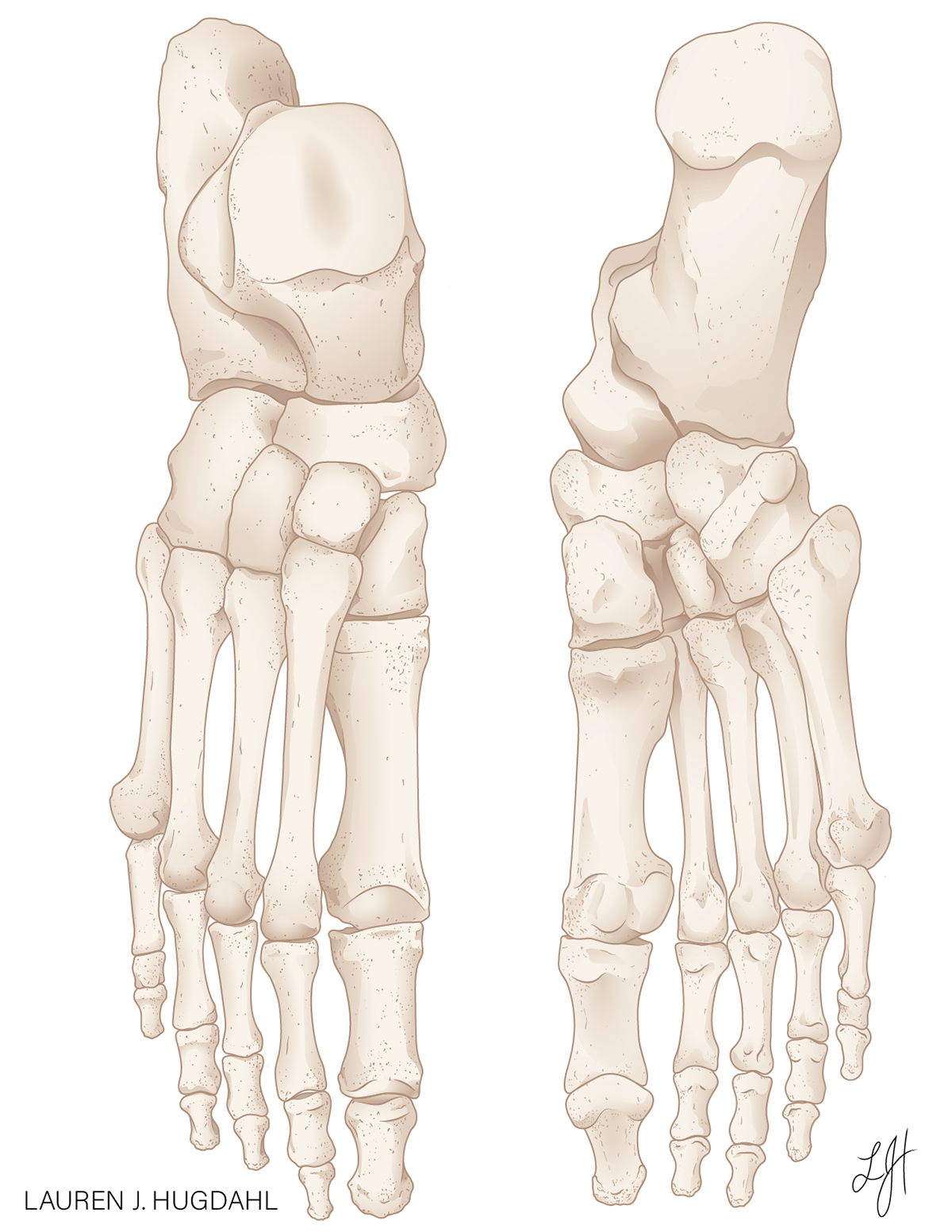
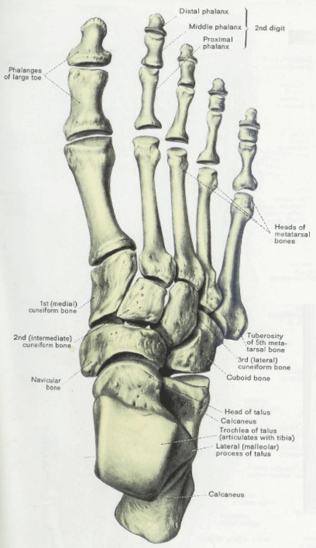
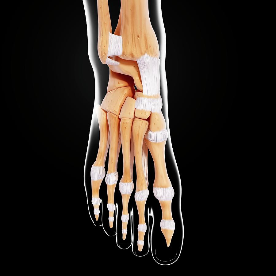
Bones of foot Bones of foot The 26 bones of the foot consist of eight distinct types, including the tarsals, metatarsals, phalanges, cuneiforms, talus, navicular, and cuboid bones. The.

Foot Bone Anatomy Vector Illustration 539973 Vector Art at Vecteezy
33,955 skeleton foot stock photos, 3D objects, vectors, and illustrations are available royalty-free. See skeleton foot stock video clips Filters All images Photos Vectors Illustrations 3D Objects Sort by Popular

Foot & Ankle Bones
2,490 Skeletal Foot Stock Photos, High-Res Pictures, and Images - Getty Images Images Creative Images Browse millions of royalty-free images and photos, available in a variety of formats and styles, including exclusive visuals you won't find anywhere else. See all creative images Trending Image Searches Happy Holidays Christmas Background Christmas
Bones and Joints of the Foot and Ankle Overview FootEducation
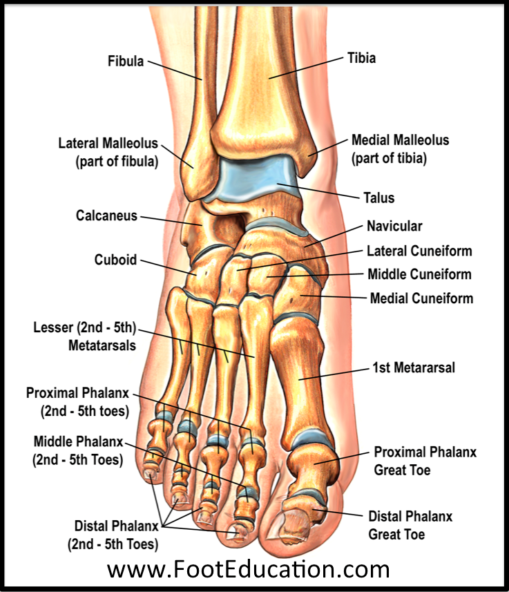
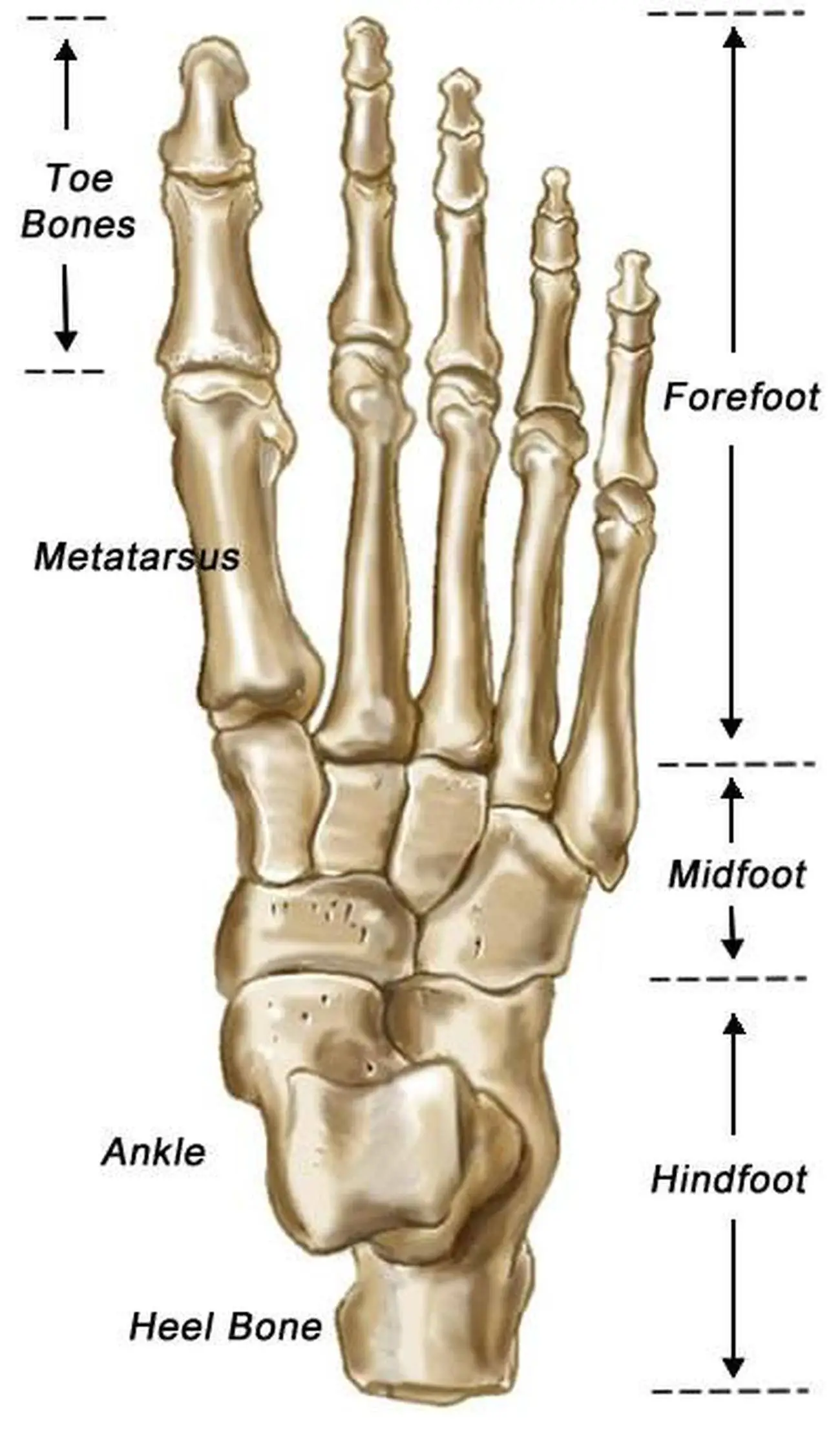
Last updated 2 Nov 2018 The anatomy of the foot The foot contains a lot of moving parts - 26 bones, 33 joints and over 100 ligaments. The foot is divided into three sections - the forefoot, the midfoot and the hindfoot. The forefoot
Foot Skeleton Drawing at GetDrawings Free download
Browse 2,627 human foot anatomy photos and images available, or start a new search to explore more photos and images. NEXT Browse Getty Images' premium collection of high-quality, authentic Human Foot Anatomy stock photos, royalty-free images, and pictures.

3D model Skeletal Foot VR / AR / lowpoly CGTrader
Human body Foot Foot The foot is the lowermost point of the human leg. The foot's shape, along with the body's natural balance-keeping systems, make humans capable of not only walking, but.
Pictures Of Bones Of The Feet
Common causes of foot pain include plantar fasciitis, bunions, flat feet, heel spurs, mallet toe, metatarsalgia, claw toe, and Morton's neuroma. If your feet hurt, there are effective ways to ease the pain. Some conditions specific to the foot can cause pain, less movement, or instability. Verywell / Alexandra Gordon.
foot skeleton 3ds
The talus is held in place by the foot bones surrounding it and various ligaments. 4. Calcaneus. The calcaneus is more commonly known as the heel bone. It is the largest of the foot bones and has a quadrangular shape. The calcaneus is the most commonly fractured tarsal bone, usually from a high fall.
Human Foot Bones Photograph by Sebastian Kaulitzki Pixels
These bones are arranged in two rows, proximal and distal. The bones in the proximal row form the hindfoot, while those in the distal row from the midfoot. Hindfoot. Talus. Calcaneus. The talus connects the foot to the rest of the leg and body through articulations with the tibia and fibula, the two long bones in the lower leg. Midfoot. Navicular.
Human Foot Bones Photograph by Pixologicstudio/science Photo Library Pixels
Details. The original file was in Wavefront .OBJ format. The following is the original legend from the file: Foot Bones # # Courtesy of: # # Viewpoint Animation Engineering # 870 West Center # Orem, Utah 84057 # (801)224-2222 # 1-800-DATASET # $ Contributed to the FTP site at avalon.chinalake.navy.mil (129.131.31.11) # by Scott R. Nelson of Sun.

Womans Foot Bones Labeled On White Stock Photo Download Image Now Human Skeleton, Ankle
Browse 9,900+ skeletal foot stock photos and images available, or start a new search to explore more stock photos and images. Sort by: Most popular Run off your heels Rear view shot of the highlighted joints in a runner's foot Valgus deformity of the big toe. Foot health care. Valgus deformity of the big toe. Foot health care. Vector illustrations.
